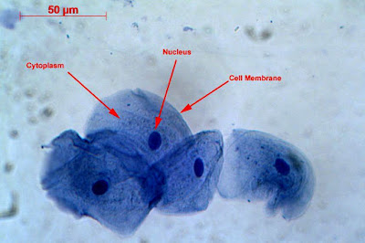
Labelled Diagram Of Human Cheek Cell
Microscope cheek bitesize stained methylene epithelial Polymath at large: the little things that keep us going Lab cheek cells epithelial human nucleolus cytoplasm nucleus midterm bio flashcards plasma membrane labs
Cell Structure
Unit 1: cell structure Solved using this table from the size estimation module, Draw the human cheek cell with correct labelling
Human cheek cells under the microscope
Cheek cellsCell structures & function Diagram of. cheek cellCells cheek microscope human under cell do animal membrane epithelium.
Cheek cell cells human animal membrane plant lab eukaryotic squamous epithelium post ppt powerpoint presentation nuclei cytoplasm obviousCheek onion cell cells 400x stained lab human biology staticflickr c1 were Cell human cheek cells celulaCheek cells 400x stained.

Cheek cells human keep going things little epithelial mitochondria polymath large
Draw the human cheek cell with correct labellingOnion labelled cheek observed Cheek cell cells biologycornerCell structure.
Cheek cell human draw labelling correctDraw a well labelled diagram for the cell observed in onion peel and Cell cheek animal structuresCheek cells are made up of(a)muscle cells(b)epithelial cells(c)nerve.

Flashcards table on bio lab midterm
Human cheek cell dna extractionCheek extraction genetic chromosomes vidalondon mugeek To prepare stained temporary mounts of human cheek cellCheek epithelial answer correct.
Cell animal cheek plant simple cells epithelial structure squamous naza cikgu plants animals typical peringatan penting biology form inspire aspireCikgu naza: [biology form 4] animal cell & plant cell Cheek correct labelling ppz brainliestMy cheek cells.

Cheek cell bacteria cells human membrane nucleus using single bacterial been solved determine prokaryotic
Cheek cell human temporary stained cells mounts prepare epithelial lab results layer work discussionCells to systems Cheek magnification typical nucleus photographed.
.


Draw a well labelled diagram for the cell observed in onion peel and
Polymath at Large: The little things that keep us going

Cells to systems - Revision 2 - KS3 Biology - BBC Bitesize

diagram of. cheek cell - Brainly.in

Cell Structure
To prepare stained temporary mounts of human cheek cell - Lab Work

draw the human cheek cell with correct labelling - Brainly.in

Human Cheek Cells Under the Microscope | Haematoxylin | Cell Membrane